Auto in situ Hybridization
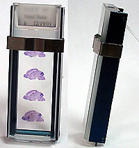
Slides carrying tissue sections are integrated into flow-through chambers (Image 1), which are then placed into a temperature-controlled rack (Image 2) positioned onto a Tecan Genesis liquid handling platform. All solutions are added using a computer-controlled liquid handling system. Temperature of the rack and solutions is controlled by water circulator baths that are regulated by the PC that controls the liquid handling system. The following main steps are sequentially carried out by the GenePaint robot in an automated manner:
If the robot is fully filled with slides (192 slides) the entire procedure requires approximately 24 hours. |
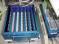 Image 2 |
|
Tissue Collection |
|
|
Mice are maintained on a 12-h light cycle with light being turned on at 07:00. Animals are anesthetized with isoflurane (Abbott Laboratories) and either killed by decapitation (for brain preparations) or through cervical dislocation (for embryo collection). Tissues are processed as follows: Method 1: Embryos are cryo-protected with sucrose and embedded in gelatin and frozen in special copper chambers (see below). Method 2: Embryos are embedded in paraffin using standard methods of paraffin embedding. E14.5 embryos, heads of E15.5 embryos: Following a brief wash in cold phosphate-buffered saline, tissue is transferred to ice-cold O.C.T 4583 (Tissue-Tek, Sakura). Tissues are transferred to a freezing chamber (Image 3) within 5 min after dissection. A freezing chamber consists of a square copper base and transparent Plexiglass side walls. The chamber is placed on a specially designed, movable stage fitted to a stereomicroscope. Through translation and rotation of this stage, the edges of the chamber can be aligned parallel to a grid placed into one of the eyepiece of the stereomicroscope. The chamber is filled with O.C.T. and placed for 40 sec onto an aluminum block submerged by 2/3 in a dry ice/methylpentane mixture (maintained at -65°C). The chamber is then removed and the tissue is submerged in O.C.T. and oriented appropriately with the aid of a blunt dissecting needle. Orientation is achieved if the dorsal midline of the embryo is parallel to the gridlines of the eyepiece and the sagittal midplane of the brain is parallel to the side walls as judged by viewing the specimen frontally through the transparent side walls. After orientation, usually within 1 min, the chamber containing the specimen is returned to the cooling device where it continues to freeze in a uniform fashion with the plane of freezing moving upward at a rate of about 3 mm per minute. Following freezing, the chamber is disassembled. O.C.T. blocks containing tissue are placed into plastic bags and are stored at -80°C for several months. Gelatin embedding of E10.5 embryos is carried out in an analogous manner. Embryos are oriented with their mid-plane parallel to the copper plate. P7 and P56 brains:The dissected brain is placed into a dish filled with ice-cold O.C.T. and shortly thereafter the tissue is placed and frozen in the freezing chamber as described above. Orientation: midline of the brain is set parallel to the grid of the eyepiece and equivalent structures of the brain are equidistant to the copper plate, as judged by viewing the brain frontally through the transparent side walls. |
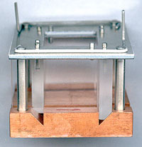 Image 3 |
|
Tissue Sectioning |
|
|
Frozen sections: At least one day prior to sectioning, O.C.T. blocks are transferred to a -20°C freezer and then mounted on a cryostat chuck. Because of the orthogonal walls of the O.C.T. block, specimens can be accurately oriented. The chuck is fastened to the goniometer of the cryostat and oriented so that the edge of the knife cuts parallel to one of the cardinal planes of the specimen. A Leica cryostat Model CM 3050S is used and sections are 20µm or 25µm thick. Superfrost slides are placed into aluminum slide holders (Image 4) which permits precise positioning of sections onto the slides. Following sectioning, slides are kept in slide racks in the cryostat until ready for fixation. Fixation, carried out in a Leica Autostainer XL, is in 4% paraformaldehyde for 20 min at room temperature. Subsequently, slides are washed for 5 min in PBS, acetylated and dried by passing them through an ethanol series ending with 100 % ethanol. After drying at 30°C, slides are stored at -80°C in boxes containing a drying agent and sealed with electrical tape. Paraffin sections: Only E10.5 embryos are paraffin embedded and they are sectioned at a thickness of 10µm. Following sectioning the tissue is deparaffinized, fixed in 4% paraformaldehyde, acetylated and dehydrated and stored in the presence of desiccant at -80°C. Gelatin sections: Tissue embedded in gelatin is sectioned in a cryostat at a thickness of 20µm and treated as described for frozen sections. |
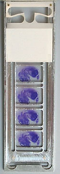 Image 4 |