Auto Microscopy
|
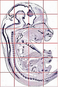
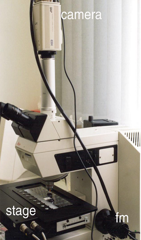
Slides generated by the in situ hybridization robot are coverslipped and photographed in a light microscope equipped with a motorized stage (Image 2) that moves the slide in front of the objective. The equipment used consists of a Leica DM-RXA2 microscope, a motorized Märzhäuser stage that accommodates up to eight slides, a Leica electronic focusing system, a Hitachi CCD camera and a PC based controller that drives stage and camera. Brain sections are too large to be photographed as a whole. Therefore, the motorized stage moves the sections in a stepwise fashion in front of the objective and at each step an image is taken. Each image is stored as a bitmap file and individual images are assembled into a mosaic image (Image 1) that is cropped, properly oriented and saved as TIFF file. Resolution of images:
|